


Osteochondrosis is a chronic disease expressed in articular cartilage by malnutritional diseases. Osteochondrosis of the spine usually occurs when the disc and intervertebral joints change. According to the location, cervical, thoracic and lumbar osteochondral diseases are distinguished. Osteochondrosis is also often found, in which all spinal components suffer. Pathology requires consultation with a doctor and comprehensive treatment methods.

describe
Like other chronic diseases, spinal diseases are rapidly "young". If earlier back and joint pain plagues older people, patients aged 18-30 are increasingly treating doctors today.
Scientists believe that a person's directness is a prerequisite for the development of this disease, and long-term sitting posture also promotes osteochondrosis. In addition, as you age, the articular cartilage will lose its elasticity and elasticity and become thinner. The intervertebral disc loses moisture and absorbs ability and becomes vulnerable when the body is tired.
reason
The cause of cervical osteochondrosis is load, which affects the cervical area under different circumstances. As a result, the muscles in the neck start to drop significantly, thus compensating for this load, thus spasms occur and violates blood flow in the area.
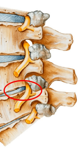
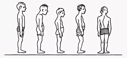
- Violation of postural scoliosis, curvature, roundness, flexion of the brain and other postural diseases, even if they are insignificant, can seriously violate the balance of the spine. As a result, the load on the intervertebral disc is distributed unevenly, which causes its deformation and increased wear. The vertebrae begins to get closer, leading to the invasion of nerve processes, and cervical osteochondral disease develops very quickly. Similar consequences also violate postures caused by changes in the natural position of the ribs.
- Muscle spasms of the back muscles, breasts, and stress can cause very tense parts of the body. As a result, the body's general balance position is disturbed, causing changes in the spinal position. Deformation may affect the cervical vertebrae area or other parts of the spine, resulting in osteocartilage degeneration in the chest, cervical vertebra and waist areas.
- Violation of blood supply, since vertebrates have no direct contact with the circulatory system, they get nutrition from the surrounding tissues. Violation of blood supply from the cervical spine leads to the fact that the disc does not receive enough fluid to rehydrate (form restored due to the absorption of water) and renewal of cartilage tissue. As a result, their wear is accelerated and the distance between the vertebrae in the cervical vertebrae region decreases, resulting in osteocartilage degeneration.
- Violating nerve behaviors, reduced sensitivity of nerve roots can lead to pathological changes in their structure, so the patient's vertebrae displacement and deformation have not yet attracted the patient's attention. After all, there is a lack of pain due to sensitivity disorders.
- Diseases of internal organs are the wrong locations of internal organs, and their displacement and reduction can lead to invasion of general balance in the body due to various dysfunctions. As a result, this acutely affects the location of the spine - cervical, lumbar spine replacement and deformation, resulting in the corresponding type of osteochondrosis.
Often, osteocartilage in the cervical area develops because the influence of adverse external factors violates the natural balance position of the spinal column and other systems of the human body.
diagnosis
The diagnosis of cervical osteochondrosis begins with the collection of all necessary information about the patient. Experts asked about the complaints that plagued the person, were interested in his professional activities, and how he spent the weekend. An important point is that parents and grandparents have osteochondriasis, because it is a genetic disease.
The doctor then directly performs a visual examination of the patient. He studied the cervical cabin and curvature the cervical area. This allows experts to assess the extent of the disease progression, as palpation of the cervical area can cause severe pain in the late stages.
When checking, you should note:
- About the severity of the cervical spine;
- height of the patient's shoulder;
- Possibility of asymmetry in the upper superloke zone;
- the possibility of neck asymmetry (e. g. , the result of congenital pathology or sharp muscle spasms);
- muscle condition of shoulder straps and upper limbs (e. g. , one aspect of muscle atrophy may indicate compression of the cervical spine);
- Chin position - The chin should be in the midline normally;
- movement of the neck (bend, tilt right, rotate and rotate).
Palpation is performed at the patient's initial position:
- Lying behind;
- Lying on the belly;
- Sit on a chair.
A study on exercise volume was also conducted. It is performed in the initial position of the patient sitting in a chair (to fix the other spine).
The following basic movements that distinguish the cervical area:
- buckling;
- Expand;
- Ramps to the right and left;
- Rotate.
About half of the flexion and stretching volume occurs on the back of the head, i. e. between the vertebrae of C1 and C2. The rest of the movement is performed due to the underlying vertebrae, with a lot of movement in the C5-C7 vertebrae. The roll angle is evenly distributed between all vertebrae.
For accurate diagnosis, other studies were conducted:
- The cervical area of the X-ray. This method is suitable in the early stages of the disease, but may not be useful in advanced forms.
- CT (Computed Tomography). This allows you to see the structural changes of the vertebrae, but with this method, it is impossible to determine the size of the hernia between the vertebrae.
- MRI. It is considered the most effective method to diagnose cervical osteochondrosis. You can determine the size of hernia between the discs and how much they develop.
- Doctors can also prescribe a double-strand scan that allows you to determine behavior that violates normal blood circulation in the artery.
treat
Treatment of cervical osteochondrosis is a complex therapy, including medication, gels, and various physical therapy measures. The important role of massage in the therapeutic gymnastics and problem areas.
drug
Without medication, it is impossible to eliminate the consequences of changes in spinal degeneration disease. Among the complex rehabilitation methods used by most traditional medical experts, the use of drugs in osteochondrosis treatment in the cervical vertebrae region is one of the main points of the complex approach.
It is designed to solve several problems, including:
- Stop pain symptoms and eliminate the inflammatory process;
- Remove muscle cramps;
- The process of stimulating the regeneration of cartilage and bone tissue cells;
- Enhance the body's protective properties and increase immunity;
- Improve general conditions by eliminating other symptoms of disturbing recovery.
According to these purposes, all medications prescribed by a physician with cervical osteochondrosis can be conditionally divided into the following groups:
- Analgesics (non-replacement medications that relieve pain).
- Anti-inflammatory (steroids) are hormone drugs that relieve inflammation and eliminate pain.
- Cartilage protectors are drugs that contain cartilage tissue components - chondroitin, hyaluronic acid.
- musorelaxant. These medications can relax muscle tone. They are used in surgery and orthopedics as auxiliary remedies to prevent pain. Such drugs are administered by parentel intestine and are therefore always performed under the supervision of a doctor.
- Vitamin. As osteocartilage degeneration in the cervical area, vitamins are prescribed, which can beneficially affect the peripheral nervous system and improve conductivity. Water - Dissolved Vitamins: B1, B6, B12, Fat - Solvent - Solute Vitamins: A, C, D, E. In recent years, drugs containing painkillers and vitamin ingredients have been combined more frequently.
- Ointments and gels are used for external purposes.

Physical therapy
The main goal of physical therapy is to stimulate the regeneration process in the body and eliminate pain. The most popular way to treat cervical vertebrae is:
- ultrasound. Ultrasound is used to relieve severe pain manifestations and inflammatory responses. Ultrasound is to massage the neck tissue, which will then metabolize.
- Vibration massage. The effect on the pain area during vibrating massage is through mechanical oscillation movement. To properly perform this physical therapy, a belt-like vibrator is usually used.
- Electrophoresis. This method of physical therapy care for cervical bone cartilage is performed using bilinear and modulated currents and when the body is administered to the drug tissue. Electrophoresis perfectly relieves spastic syndrome and eliminates inflammation of muscle pain.
- Magnetic therapy. The essence of magnetic therapy as physicalization of osteochondrosis in the cervical region is explained by the use of constants or variables of magnets (frequency of different sizes). This approach can help patients eliminate pain and stop the inflammatory process in the furnace. After purchasing special magnetometer equipment, the process is usually done at home.
- Dutoenzor-therapy. Currently, a fairly popular method of physical therapy involves stretching the spine in a patient’s body. To perform such a procedure, mattresses must be arranged in a special way, which tilt the ribs, which change positions under the weight of their own body. Muscle tone is normalized, resulting in their relaxation.
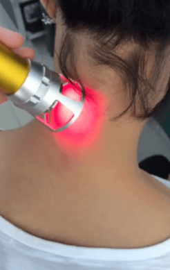
- Laser therapy. Lasers have complex effects on the focus of inflammation, which activates biological processes in nervous system tissues. This allows you to get a positive impact from treatment. Complex effects on the body are anti-inflammatory, analgesic and wound healing effects. A laser treatment procedure must not exceed 15 minutes. This is the best time for a spiral neon laser to be on the affected area. In this case, the laser duration should not exceed 2 minutes.
- Balneotherapy. The benefits of mineral water have been a long time, and that's what Balneotherapy is based on. This procedure means actively using water resources to treat osteochondrosis. In addition to adopting a bathtub, different types of souls and active swimming in the pool, the therapy involves applying therapeutic mud to areas of the body’s pain. The therapeutic effect is achieved by the simultaneous action of chemically active substances contained in water under various temperature conditions. This technology allows you to stop pain syndrome by improving local microcirculation in your tissues.
Exercise therapy
It should be remembered that when signs of aggravation begin to appear, no exercise therapy is performed: pain. After the LFK complex, they can exacerbate and cause inconvenience.
There are many general suggestions for charges:
- Sports should be done in a well-ventilated room, which is a great choice for the street.
- The course is only performed during the disease remission period (without symptoms).
- Clothes in exercise therapy should be wide, rather than embarrassing movements and breathing.
- All movements are smooth, with the magnitude and number of repetitions gradually increasing.
- If the pain begins, you should stop the course immediately.
- Finish the measurement of pressure and pulses before the category. When these indicators are different from normal conditions, the load should be reduced.
- It is recommended to listen to your breath throughout the course, which will increase efficiency. All stretching exercises are performed while exhaling.
- It is important to gradually increase the number of loads and repetitions, which will reduce the risk of injury and prevent overwork.
- Exercises are important for regular execution, so you can achieve fast results.
- Before you start an independent course, you need to consult your doctor and agree to his series of exercises.
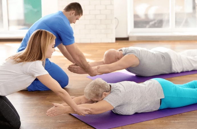
Recommended exercises are located at the start of the stomach:
- The end of the head is on the forehead, the hands are on the back of the head, and the elbows are parallel to the floor. Lift your head from the floor with both hands and keep this position in 4 accounts and relax. Repeat 2-4 times.
- The head stops on the chin, the palms under the chin. In time, stretch your arms forward, two - stretch forward, four - start position. Repeat 2-4 times.
- The hand stretched forward. Swimming "rabbit" style, repeat 4-8 times.
- The palm under the chin emphasizes the palm of the forehead. Or, take out the heel of the hips. Repeat 4-8 times.
Recommended exercises are located at the starting position on the side (right, then left):
- With your right hand out, your right ear lie on it, lift your right hand with your head, fix the position to 4 accounts, lower and relax. Repeat 2-4 times.
- Place your left hand on the floor in front of your chest and your left leg moves the fly back and forth. Repeat 6-8 times.
- Lift your left hand along your body, lower, lower. Repeat 2-4 times.
- Left hand on thigh. Pull both knees to your chest while exhale and stretch your legs inspire. Repeat the exercises 2-4 times.